-
YAG Laser
-
Specular Microscope
-
Contrast Sensitivity Testing Device
-
Electroretinogram (ERG)
-
Laser Flare Meter
-
Fundus Camera
-
Optical Coherence Tomography (OCT)
-
Ultra-widefield Retinal Imaging Device
-
Angiography Device
-
Visual Field Analyser
-
Anterior Segment Ophthalmic OCT Device
-
Ophthalmologic Ultrasound (A/B Scan)
-
PASCAL Laser
-
IPL Laser for Dry Eye Treatment
YAG (Yttrium Aluminum Garnet) Laser is a device that utilizes lower yet efficient energy compared to traditional laser equipment for treatment. It is commonly used for laser treatment, particularly in treating posterior capsule opacification (PCO), which is the most common complication of cataract and glaucoma surgery.
-
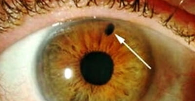
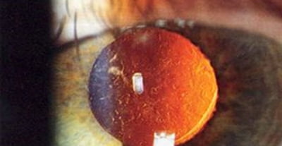
Laser Synechiolysis
-
PCO after Cataract Surgery
(Laser Capsulotomy / Source: Wikipedia)
Example of YAG Laser Treatment Process
YAG Laser is set up → Patient’s mydriasis is checked → Anesthetic is instilled into the patient's eyes → Patient is seated and laser treatment is performed
The cornea—which is only about 0.5mm thick on average—is composed of five layers, and among them, the innermost layer called the endothelium is crucial for the overall health of the cornea. Specular Microscope plays a significant role in examining the health of the corneal endothelium, from examinations before and after ICL (implantable contact lens) to examinations for corneal edema.


Healthy endothelial cells in a 30-year-old male are characterized by the following
- Approximately 2500 to 3000 arrays per square millimeter
- A similarity rate of 60% or more in the shape of cells, resembling a regular hexagon
Contrast Sensitivity Testing Device utilizes precisely controlled sine-wave gratings via a computer to accurately measure visual function. It provides precise information on visual function before and after surgery, serving as a crucial indicator for assessing improved conditions post-surgery.
- R
- L

- Measured contrast sensitivity of the patient
- Normal objects as perceived by the patient
- Objects as perceived by the patient reflecting the measured contrast sensitivity
Electroretinogram (ERG) measures intricate details regarding the condition, progression, and state of the retina by analyzing electrical signals produced by retinal nerve cells when exposed to light. ERG examinations are especially beneficial for evaluating patients prior to cataract surgery and for the diagnosis of retinal conditions or glaucoma. At The One Seoul Eye Clinic, the entire ERG procedure—including preparation and examination—takes about 7 minutes in total, and the electrodes used for the examination are non-intrusive, ensuring optimal comfort for patients
-

Example of Measurement Results:
Right Eye Normal / Left Eye Abnormal -

Non-invasive Electrodes
Laser Flare Meter is a device that measures the presence and condition of intraocular inflammation (uveitis, keratitis, scleritis, etc.) based on the number of protein clusters within the aqueous humor, reflected and confirmed through the principle of light scattering after projecting a laser into the eye. This equipment is useful for outpatient examinations and follow-ups after cataract surgery.
-

Front View
-

Side View

Example of Measurement Results
In a normal eye, an average of 2.9 to 3.9 photons is observed per millisecond. If more than 30 photons are observed per millisecond, it is considered indicative of inflammation.
Fundus Camera is the first truecolor confocal scanner that accurately recreates the true colors of the retina. It is capable of capturing images automatically even with a small pupil, providing high-resolution images of the retina and optic nerve
-

Macular Degeneration Detected by Fundus Camera
-

Central Retinal Vein Occlusion Detected by Fundus Camera
Optical Coherence Tomography (OCT) is essential for the diagnosis and treatment of various retinal conditions such as macular degeneration, diabetic macular edema, epiretinal membrane, and macular hole. Through the vascular imaging mode, it enables non-invasive and more precise examination of subtle vascular changes that were not detectable with conventional equipment
-

Epiretinal Membrane Detected by OCT
-

Macular Hole Detected by OCT
-

Glaucoma Detected by OCT
The Ultra-widefield Retinal Imaging Device is an advanced retinal imaging equipment that can capture images of the peripheral retina without dilation. Utilizing the wide-field autofluorescence mode facilitates easy diagnosis and assessment of the progression of macular degeneration. Additionally, the wide-field fluorescein angiography mode allows the diagnosis of vascular changes in the peripheral retina that were previously undetectable with conventional fluorescein angiography
-

Retinal Detachment Detected by Ultra-widefield Retinal Imaging Device
-

Diabetic Retinopathy Detected by Ultra-widefield Retinal Imaging Device
Through the autofluorescence mode, the diagnosis and progression of dry macular degeneration can be assessed. Using indocyanine green angiography (ICG), choroidal neovascularization can be identified, and with fluorescein angiography (FAG), retinal neovascularization can be detected.
-

Macular Degeneration Detected
by Autofluorescence Mode -

Macular Degeneration Detected
by Indocyanine Green Angiography
Visual Field Analyser is a device used to examine narrow-angle glaucoma, utilizing the built-in software of the analyzer to automatically diagnose the progression of glaucoma.
-

Visual Field Test Results of
a Glaucoma Patient -

Screen displaying automatic diagnosis results regarding
the progression of glaucoma using the built-in
software of the analyzer
(GPA, Glaucoma Progression Analysis)
Anterior Segment Ophthalmic OCT Device is a versatile equipment used for various purposes, including measuring axial length and calculating intraocular lens power during cataract surgery, as well as capturing images of the anterior segment for glaucoma screening. Previously, separate tests such as corneal topography, ocular biometry, and gonioscopy were required, but with this device, one can obtain high-precision images and comprehensive measurements in a single examination, significantly reducing the need for multiple tests.
-

Corneal Topography Test Results of a Keratoconus Patient
-

Imaging Results Capturing the Narrowed Anterior Segment Due to Progressing Cataracts
Ophthalmologic Ultrasound (A/B Scan) is essential equipment used to determine the presence and degree of retinal detachment when it is difficult to observe through fundus examination, and to diagnose or differentiate tumors.
-

Retinal Detachment Detected by Ultrasound
-

Choroidal Tumor Detected by Ultrasound
PASCAL Laser features enhanced functionality compared to traditional lasers, providing less pain during treatment and significantly reducing treatment time. It is commonly used for laser treatments in conditions such as retinal break, diabetic retinopathy, and glaucoma
-

Screen displaying selectable laser patterns
according to the situation to reduce treatment time -

Performing Panretinal Photocoagulation
(PRP) with PASCAL Laser
IPL (Intense Pulsed Light) laser is used to treat dry eye syndrome caused by meibomian gland dysfunction, which are oil glands along the rims of the eyelid inside the tarsal plate. The equipment irradiates the IPL laser to destroy abnormal blood vessels and heat to melt and open clogged meibomian glands

Put on eye protection shield → Apply gel to the treatment area → Irradiate M22 IPL laser (approximately 5 minutes) → Remove gel and cool & soothe if necessary (5 minutes)
-

Inflamed and Hardened Oil Glands Before IPL Treatment
-

Clear Oil Glands After IPL Treatment
-
Weekdays
08:30 a.m. - 05:30 p.m -
Saturdays
08:30 a.m. - 01:30 p.m -
Lunchtime
01:00 p.m. - 02:00 p.m
8F and 9F Sinsa Square, 652 Gangnam-daero, Gangnam-gu, Seoul
o straight for 5 minutes (330m) on foot from
Exit 6 of Sinsa Station on Subway Line 3